Welcome: GUANG DONG ASCEN TECHNOLOGY CO.,LTD
Language:
 ∷
∷

∷
∷
Sperm Morphology
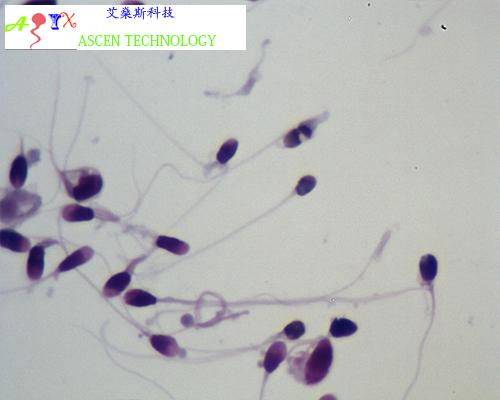
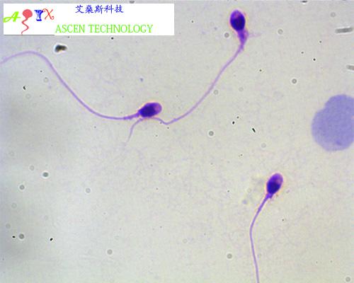
Morphology of the sperm head is an important criterion for the correct diagnosis. The software is set up to analyze still images of smears stained with the Diff-Quik stain according to strict Krueger’s criteria. We have selected Diff-Quik as worldwide recognized leader in rapid staining of sperm. With Diff-Quik, the head is stained pale blue in the acrosomal region and dark blue in the post-acrosomal region which is a good basis for precise image analysis. The following parameters are assessed for every spermatozoon:
§ Area of the head.
§ FFC = form factor circle. The degree of similarity of the sperm head to a circle.
§ Perimeter of the head
§ Brightness.
§ ELL_B = Big axis of ellipse outlining the sperm head, the length of the sperm head.
§ ELL _S = Small axis of ellipse outlining the sperm head, the width of the sperm head.
§ Elng = elongation of the sperm head.
§ FFE = form factor ellipse. The degree of similarity of the sperm head to an ellipse.
§ Acrosome = Percentage of the acrosomal region.
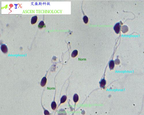
The software classifies spermatozoa into Norm and Head Pathology classes automatically based on head parameters. You c can easily correct the results manually and also specify other anomalies (Tail Pathology, Neck Pathology). An extended morphology classifier is available which allows you to specify the following spermatozoa structure abnormalities indicating potential infertility: tapered, pyriform, round, amorphous, vacuolated, small acrosome, double head, pinhead, bent neck, asymmetrical neck, thick insertion, thin neck, short tail, bent tail, coiled tail, excess residual cytoplasm (ERC).
In case you can not receive a good image of stained smear suitable for automated detection (e.g. because of incorrect sample preparation), there is a special tool which allows you to outline the cells manually.
Software is able to detect both samples prepared according to WHO recommendations for CASA including centrifugation (recommended) but also can be adjusted to easier procedures which provide higher background staining. For complicated cases, there is an option of manual drawing and deleting of objects.
It is now customary to record the number of morphological sperm defects divided by the number of defective spermatozoa, a measure called the teratozoospermia index (TZI). After the analysis is finished, the TZI is calculated automatically. The teratozoospermic index values should read between 1.00 (each abnormal spermatozoon has only one defect) to 3.00 (each abnormal spermatozoon has head, neck and tail defects).
Contact: Alex Chan
Phone: 13823571046
Tel: 0759-87966
Email: info@szyessmt.com
Add: E Building, Zi Jing Garden,Bi Feng Tang,Hong Wu Road,Xiashan District,Zhanjiang City,Guangdong Province,China